| Magyar verzió |
|||
| News | |||
|
|||
| Standard | |||
| Origin, character | |||
| Articles, infos | |||
| Photo album | |||
|
|||
| Breeders | |||
| Puppies | |||
| Stud dogs | |||
| Genetic diseases | |||
|
|||
| Show results | |||
| Show schedules | |||
| Show photos | |||
| Champion titles | |||
|
|||
| Links | |||
| Guestbook | |||
| Contact | |||
| GENETIC PROBLEMS OF THE TIBETAN TERRIER |
| INFO | |||||||||||||||||||||||||||||||||
The Tibetan is a very hardy breed and is considered long-lived with most living well beyond 12 years and many to 15 or 16 years. Some problems found in the Tibetan Terrier are: hip dysplasia (HD), patella luxation (PL), progressive retinal atrophy (PRA), lens luxation (LL), hypo-thyroidism, cataracts and canine neuronal ceroid lipofuscinosis (CCL/NCL). Some of these problems have been proven to be hereditary, and conscientious breeders have screened their stock and can explain these problems and their incidence. Many breeders will have knowledge of their puppies’ bloodlines and potential buyers are advised to ask questions and inquire as to evidence of any testing done on the sire and dam. Click on the problem's name to read about it. |
|||||||||||||||||||||||||||||||||
|
|||||||||||||||||||||||||||||||||
| HIP DYSPLASIA | |||||||||||||||||||||||||||||||||
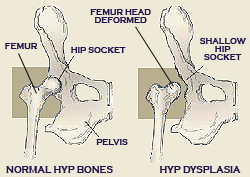
Hip Dysplasia is a terrible genetic disease because of the various degrees of arthritis (also called degenerative joint disease, arthrosis, osteoarthrosis) it can eventually produce, leading to pain and debilitation. The very first step in the development of arthritis is articular cartilage (the type of cartilage lining the joint) damage due to the inherited bad biomechanics of an abnormally developed hip joint. Traumatic articular fracture through the joint surface is another way cartilage is damaged. With cartilage damage, lots of degradative enzymes are released into the joint. These enzymes degrade and decrease the synthesis of important constituent molecules that form hyaline cartilage called proteoglycans. No one can predict when or even if a dysplastic dog will start showing clinical signs of lameness due to pain. There are multiple environmental factors such as caloric intake, level of exercise, and weather that can affect the severity of clinical signs and phenotypic expression (radiographic changes). There is no rhyme or reason to the severity of radiographic changes correlated with the clinical findings. There are a number of dysplastic dogs with severe arthritis that run, jump, and play as if nothing is wrong and some dogs with barely any arthritic radiographic changes that are severely lame. The phenotypic evaluation of hips done falls into different categories:
source: OFA |
|||||||||||||||||||||||||||||||||
 This causes the cartilage to lose its thickness and elasticity, which are important in absorbing mechanical loads placed across the joint during movement. Eventually, more debris and enzymes spill into the joint fluid and destroy molecules called glycosaminoglycan and hyaluronate which are important precursors that form the cartilage proteoglycans. The joint's lubrication and ability to block inflammatory cells are lost and the debris-tainted joint fluid loses its ability to properly nourish the cartilage through impairment of nutrient-waste exchange across the joint cartilage cells. The damage then spreads to the synovial membrane lining the joint capsule and more degradative enzymes and inflammatory cells stream into the joint. Full thickness loss of cartilage allows the synovial fluid to contact nerve endings in the subchondral bone, resulting in pain. In an attempt to stabilize the joint to decrease the pain, the animal's body produces new bone at the edges of the joint surface, joint capsule, ligament and muscle attachments (bone spurs). The joint capsule also eventually thickens and the joint's range of motion decreases.